28 October 2024
.Li Jiang, Fangting Zuo, Yuanyuan Pan, Ruilong Li, Yajie Shi, Xinyi Huang, Dasheng Zhang, Yingping Zhuang, Yuzheng Zhao, Qiuning Lin, Yi Yang, Linyong Zhu, Xianjun Chen
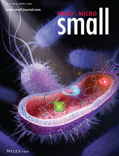
The cover of this issue is “Bright and Stable Cyan Fluorescent RNA Enables Multicolor RNA Imaging in Live Escherichia coli” by Prof. Xianjun Chen from East China University of Science and Technology, and Prof. Linyong Zhu from East China University of Science and Technology/Shanghai Jiaotong University.

Research Background
- In cell and molecular biology studies, real-time, dynamic observation of the behavior and distribution of intracellular RNAs is essential for understanding the regulation of gene expression, cellular functions, and mechanisms of disease genesis. Fluorescent RNA (FRs) technology, a powerful tool that has emerged in recent years, offers unprecedented opportunities for visualizing RNAs in living cells.FRs are a special class of RNA aptamers that bind to and activate their corresponding small-molecule fluorescent dyes, thereby generating detectable fluorescent signals within the cell. The emergence of this technology has greatly advanced the field of RNA imaging, enabling researchers to image RNA with high resolution and sensitivity in a living cell environment under near-physiological conditions.
- Although a variety of FRs have been developed and their color palette (i.e., the range of fluorescence colors that can be emitted) has been significantly expanded, cyan (cyan) FRs, which are bright and stable, biocompatible, and orthogonal to existing FRs, remain an unmet need. Cyan fluorescence offers unique advantages in biological imaging, such as lower cellular background fluorescence interference, longer fluorescence lifetime, and ease of multiplex imaging with other color channels. Therefore, the development of a novel cyan FR is of great significance in promoting the further development of RNA imaging technology.
Research Implications
- This study successfully developed an RNA aptamer named Myosotis, which can specifically bind and activate a novel GFP-like chromophore DBT to emit bright cyan fluorescence. This discovery not only enriches the color palette of FRs but also provides a new tool for RNA imaging.
- The high affinity (nanomolar level) of Myosotis for DBT and its weak dependence on magnesium ions for the folding process allows it to maintain stable performance in different cellular environments. In addition, the Myosotis-DBT complex has a long fluorescence lifetime, good photostability, and enhanced cellular brightness, properties that make it an ideal probe for RNA imaging.Myosotis-DBT exhibits good orthogonality with existing Pepper and Clivia FRs, meaning that they can be used simultaneously in the same cell without mutually interfering with each other. This property opens up the possibility of multiplexed fluorescence imaging, allowing researchers to simultaneously observe the behavior and distribution of multiple RNAs in the same experiment.
Research Outlook
- As Myosotis-DBT technology continues to mature and improve, it is expected to be widely used in various biological systems and cell types to study the role of RNAs in the regulation of gene expression, cell differentiation, and disease genesis. Combining Myosotis-DBT with other colors of FRs, more sophisticated and fine-grained multiple fluorescence imaging techniques can be developed to enable simultaneous imaging and dynamic tracking of multiple RNAs and proteins in cells.
- Given the important role of RNAs in disease onset and progression, Myosotis-DBT technology is expected to be applied in clinical diagnosis and therapy. For example, by detecting changes in the expression level or distribution of specific RNAs, early diagnosis and prognostic assessment of diseases can be realized; meanwhile, by using RNA interference technology combined with Myosotis-DBT imaging, the effect and mechanism of RNA-targeted therapy can be monitored in real-time.
Cover Design Process
The cover design aims to highlight the theme of the nano and micro world, showcasing the innovative content of the scientific research paper on multicolor RNA imaging in living bacteria. Through visual impact and detailed representation, it captures readers' attention and conveys the core research value of the article.
The cover primarily employs a dark-toned background to accentuate the purple and red bacterial structures. The choice of purple and red not only reflects the complexity and vitality of biological systems but also creates a striking visual contrast that draws the eye. The design adopts a futuristic and sci-fi-inspired style, meticulously modeling the microscopic world of bacteria to emphasize the research characteristics of the nano and micro fields.
The modeling of the bacteria and their internal structures is highly detailed, revealing the complexity and intricacies of cellular interiors. The bacteria on the cover exhibit a lifelike appearance, with their spiky surface structures and intricate internal components rendered with precision. The RNA imaging mechanism inside the bacteria is illustrated through multicolored light effects and dynamic curves, highlighting the innovation of multicolor RNA imaging. Ultimately, this cover received high praise from the supervising professor and journal editors, leading to its successful publication!

 +86 17354022649
 service@sondii.com


 IPv6 network supported